
Mastering Melanoma
When a young, skilled dermatological surgeon asks if you mind “talking shop” with his assistant while he takes out a squamous-cell skin cancer from your forehead, you answer, “No, I don’t mind.” Especially if you’re a writer and the doctor took out a melanoma on your forearm just nine months before.
When that doctor, answers “chicken breasts and pigs’ feet” to the question of “what do dermatologists practice on in residency?”, that writer has a story that should be told.
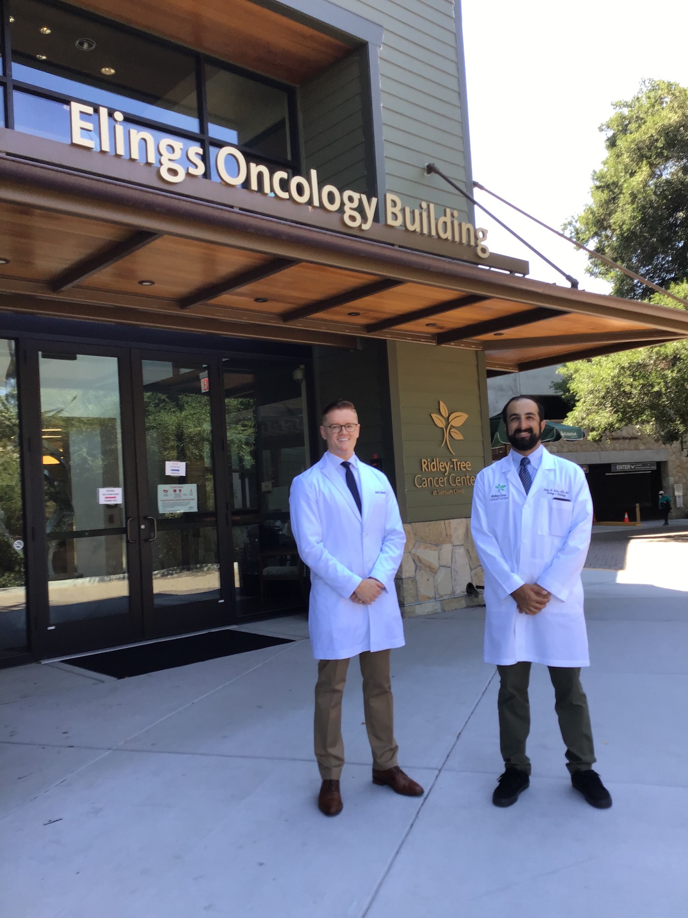
The doctor is Mark Burnett, a 38-year-old Santa Barbaran.
Doctor Burnett and his colleague, Dr. Julian Davis, an oncology cancer specialist, recently gave a lecture to the community (virtually) on how to spot and treat skin cancer as part of the Ridley-Tree Cancer Center Lecture Series. The lecture’s emphasis was on melanoma, the deadliest form, which is seeing a rise in cases here in Santa Barbara and around the country. Both doctors are on the staff of Cottage Hospital.
Born and raised in Santa Barbara, Dr. Burnett is a board-certified dermatologist and fellowship-trained Mohs micrographic surgeon at the Santa Barbara Skin Institute. Dr. Julian Davis is a Medical Oncologist at the Ridley-Tree Cancer Center. He is a published oncology researcher, and has a special interest in hematologic malignancies and advanced cutaneous cancers like melanoma.
The good news I got from the eminent doctors’ lecture is that detecting melanoma has evolved to such a degree over the past 15 years that most melanomas are now caught at their earliest stage. Innovations in Mohs surgery have vastly improved surgical cure rates and advancements in systemic medications means that even those with Stage 4 melanoma have dramatically better outcomes.
The bad news is that many people still don’t take the necessary precautions to prevent skin cancer and that there are many doctors who do not have access to the training required for recognizing the early stages of melanoma or who are not yet aware of the revolutionary new detection methods and medical treatments available.
Though the causes of skin cancer have been known, or at least suspected, for decades, what is not generally known is why cases of melanoma have increased significantly here in the U.S. Ultraviolet (UV) radiation is thought to be one of the primary drivers of this recent trend. The WHO classifies UV radiation and tanning beds as “Group 1” in carcinogens along with cigarettes and plutonium. The risk of melanoma increases by 35 percent in those who have used a tanning bed 10 or more times in their lifetime and jumps to 75 percent for those who used them before the age of 35.
People over 50, like myself, who as teenagers and young adults didn’t take sun exposure seriously and bought into the lore that a tan made you more attractive, are partially responsible for the increase in melanoma incidence. I never used a tanning bed, but I was a keen sailor, hiker, and skier.
Mohs surgery is a method that uses microscopically controlled layers around a skin cancer to ensure complete removal of all cancer cells, which can look like nothing more than a tiny, odd-looking white pimple to the naked eye (as was my case). While many dermatologists learn Mohs surgery in their residencies, only a handful are selected each year to undertake a one or two-year dedicated fellowship after their dermatology training. Dr. Burnett says, “Mohs surgery provides the highest cure rates for skin cancer because 100 percent of the surgical margin around the entire tumor is examined by the Mohs surgeon.”
More Mohs
When my first squamous cell cancers were diagnosed in New York over two decades ago, there was only one Mohs surgeon in Manhattan. The cancer had spread locally but significantly. I was fortunate that it could be excised with Mohs surgery, but I was left with a noticeable scar over my left eyebrow. For the second squamous removal, I underwent Mohs surgery and was referred to a gifted oculoplastic surgeon who repaired the surgery on my lower eyelid in his office. I lost my eyelashes, but there is no scar.
Dr. Burnett believes that, “Mohs surgery fellowships have come a long way in the last twenty years and fellowship-trained surgeons are among the most skilled at facial reconstruction following Mohs surgery, because that is all we do each day.”
He adds, however, “There are only about 1,500 fellowship-trained Mohs surgeons in the entire country and among this group only a small group of fellowships provide training to treat melanoma with Mohs surgery, which is more complex, technically, to treat than other skin cancers”.

I can attest to the evolution in Mohs surgery. The procedure performed by Dr. Burnett to remove my third squamous cell carcinoma resulted in a hole nearly one-inch in diameter in my forehead, which he reconstructed with an imperceptible outcome, as if nothing ever happened there.
Dr. Burnett says that 90 percent of melanomas do not arise from known genetic mutations, but are from sporadic mutations as a result of environmental factors such as excessive UV radiation, or increased susceptibility to UV radiation that comes with having traits such as fair skin that burns easily, red or blond hair and freckles. “However,” he says, “melanoma can appear in those who have no known risk factors, indicating that there is still a lot we do not know about the pathogenesis and development of melanoma.”
The goal of the dermatologist, Dr. Burnett says, is to detect melanoma in its early stages so that only surgery is required to excise the cancer. He emphasizes that avoiding unnecessary and excessive biopsies is important and thinks patients shouldn’t become “pincushions.”
In the past, dermatologists had only their unaided eyes to rely on in order to detect melanoma. Dr. Burnett says a doctor’s eyes are still an important factor in detection. As an example, he gives the “ugly duckling sign” that dermatologists use to distinguish when a mole looks different from other moles, as well as the ABCDE mnemonic (asymmetry, irregular border, color, and diameter of more than six mm, as well as evolving new characteristics, such as bleeding or itching).
Better Detection Through Technology
One of the leading technological advances has been the dermatoscope. Dr. Burnett compares it to the stethoscope in how it has advanced medicine for the field of dermatology. Now a standard part of dermatology training, it allows the doctor to evaluate a suspect melanoma by using specific algorithms.
Another new technology is Total Body Photography, wherein sequential photos over time are taken of the entire skin surface and then compared to new lesions. TBP has replaced memory, which was what doctors and patients previously relied upon to assess a change in a mole. When combined with a dermatoscope, the chances of detecting melanoma as early as possible rise significantly.
Pigmented Lesion Analysis (PLA) is another non-invasive tool that uses an adhesive to gently take off the skin cells overlying a suspicious mole in order to analyze the genes. This promising new technique aims to reduce the number of biopsies.
“Coming down the pipeline,” says Dr. Burnett, “is wider use of 3D Total Body Photography, which only a few academic centers have – 3D offers greater detection than 2D.” He adds that “A.I., relying on software algorithms, is also in the pipeline, but we are not at the point when a camera or robot can come in and do the scan by itself.”
All of these methods of detecting melanoma rely, in the end, on the gold standard – traditional microscopy and a trained dermatopathologist to determine if melanoma is “in situ” or “invasive.”
“In situ” is used when the cancerous cells are limited to the very top layer of the skin, the epidermis. In melanoma, that means that the cancer cells have a near zero chance of traveling to another place in the body. Most melanoma in the U.S. is diagnosed as “in situ,” a testament to advances in detection technology. Ninety-nine percent of those with “in situ” go on to live long and healthy lives.
“The likelihood of the spread of an ‘in situ’ tumor is very rare and only occurs if there was an invasive component that went undetected when the tissue was sampled,” explains Dr. Burnett. “On the other hand, an invasive melanoma has the potential to grow and thicken to such an extent that it might reach blood vessels or lymphatic channels requiring extensive surgery as well as other treatments.”
Changing Behaviors
During his part of the lecture, Dr. Burnett emphasized that behavior must change in protecting against skin cancers, especially in teenage years and the young twenties: sun-protective clothing, seeking shade, using sunscreens, and doing regular minute-long self-examination with a long and hand mirror for patients with a history of melanoma.
He also suggests taking monthly selfies of hard-to-view areas, like one’s back, in order to detect any changes from month-to-month. He says that whatever sunscreen one chooses, it should have at least a SPF of 30, be applied thick and often.
For his part, Dr. Davis said he’s “excited” to be leading, along with Dr. Burnett, the developing Santa Barbara Multidisciplinary Cutaneous Oncology Program, which also involves other local dermatologists, pathologists, surgical oncologists, radiologists, radiation oncologists, and medical oncologists. The goal of this disease-focused group is to apply and contribute to the already significant advances in treating melanoma, squamous, and other advanced skin cancers that have taken place over the past 10 to 15 years and to reduce the five thousand yearly deaths from skin cancer in the U.S.
His job, he says, is to design a treatment plan after melanoma diagnosis, based on what stage cancer a patient has, as well as the patient’s age and overall health status.
One technique used to help stage a patient’s melanoma is a sentinel node biopsy, which is performed by surgical oncologists. For this procedure, a radioactive dye and blue dye are injected into the melanoma. The dye tracks the first lymph nodes to where a melanoma would spread. Those nodes can then be removed, dissected and analyzed to see if the melanoma has spread.
The Stages
The stages of melanoma were updated in 2018 and now are: Stage 1: thin melanomas with generally excellent prognosis. Stage 2: thicker or ulcerated, but is not yet into the lymph nodes and capable of metastasizing. Stage 3: Lymph nodes are positive for cancer but the cancer has not yet spread to bones, brain, liver or lungs. Stage 4: Spread to a distant site or organ and, in general, not curable.
The classical treatment of chemotherapy has been beneficial, but with the significant toxicity of chemotherapy the treatment has had to be reevaluated. “We are very fortunate,” says Dr. Davis, “that there has been a dramatic improvement in medication to treat advanced melanoma. Instead of traditional chemotherapy, targeted molecular therapy that can block melanoma pathways, and immunotherapy, which activates the patient’s own immune system to fight their cancer, are the two main types of therapy that have shown incredible success in this disease.
The Nobel Prize in Medicine was recently awarded for this type of new immunotherapy. Dr. Davis is opening up new clinical trials for melanoma patients, which can give patients access to new cutting-edge treatments or combinations not yet approved by the FDA.
“Cancer,” says Dr. Davis, “is very good at invading the immune system. When it succeeds, it is able to shutdown the lymphocytes that are fighting back. Before this new generation of immunotherapy, survival from Stage 4 melanoma was six-to-twelve months. Now, by using a combination of drugs, five-year survival is fifty percent. We are also seeing established treatments that have proved successful in metastatic Stage 4 melanoma now filtering down to Stage 3 melanoma.”
During his lecture, Dr. Davis puts up slides of a male patient with melanoma on his scalp. The cancer was excised, but further surgery was not considered when the melanoma resurfaced as a very large skin graft would be needed. With a combination of two immunotherapy drugs, in six to seven months, the cancer was completely gone and Dr. Davis said he was able to stop the treatment.
This case was of particular interest to me, as the latest squamous cell on my forehead went into the hairline. I had asked Dr. Burnett if I was at risk of having melanoma spread to my scalp. He reassured me that scalp cancers rarely appeared in women.
I was able to offer the doctors two personal tips: The first was from the talented plastic surgeon in Manhattan two decades ago. If a combination of over-the-counter arnica and bromelain are taken 10 days before surgery, bruising will be much reduced. The second is from a beekeeping friend in Montecito: Bee Magic, a cream made from bee pollen, royal jelly, and propolis will work like magic to reduce scarring when rubbed on new and recent scars.
Answer to why chicken breasts and pigs’ feet? Because they are cheap and portable and most like human skin.
You might also be interested in...