Microscope Offers Inspiration at the Atomic Scale
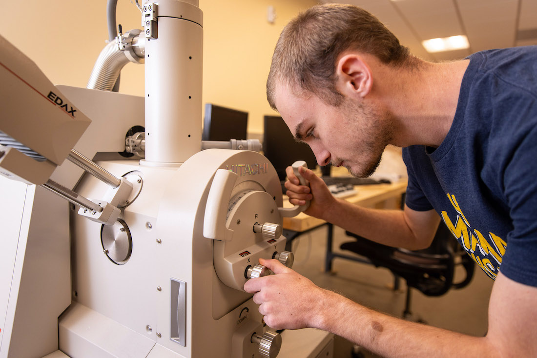
Westmont’s science professors have obtained a new tool for their teaching and research. The biology, chemistry, engineering, and physics departments will all use the Hitachi scanning electron microscope (SEM), housed in Winter Hall. Its focused beam of electrons interacts with atoms in the sample to produce an image.
Ben Carlson, assistant professor of physics, employs a Large Hadron Collider (LHC) in his research. He plans to use the microscope in his classroom as a mini particle accelerator. “It’s not directly related to my research — the energy in the electron beam is millions of times lower than in the LHC beam — but it’s a fantastic tool for teaching and doing some research,” he says. “It allows us to see things not normally visible with light because the wavelength of the electron beam is less than that of visible light. We can also measure the material composition with an x-ray detector.”
Beth Horvath, associate professor of biology, is one of the few researchers working on the taxonomy of gorgonians corals in the eastern North Pacific. She can determine the species by extracting sclerites, small calcitic skeletal bits, from the corals’ soft tissue and examining their sizes, shapes, and external surfaces. “When describing new species, it’s essential to not only provide light microscopy images of these sclerites but SEM images as well,” she says. “I’ve had to rely on an outside source to help with the SEM work. We hope this instrument will allow me and my research students to obtain SEM imagery in a timelier manner and complete journal articles faster.”
Kristi Lazar Cantrell, professor of chemistry, has focused her research on protein aggregation, including alpha-helical and beta-sheet fibril assembly. “The electron microscope will provide an opportunity for my research group to capture images of protein aggregates observed in diseases such as Alzheimer’s and Lou Gehrig’s,” she says.
Carlson says he will likely use the microscope to characterize materials, look at gold nano-particles made by the chemists, and a whole range of things they haven’t discovered. “It’s easy to use and flexible, so students have begun working with it, computing the material composition in a calibration sample.”